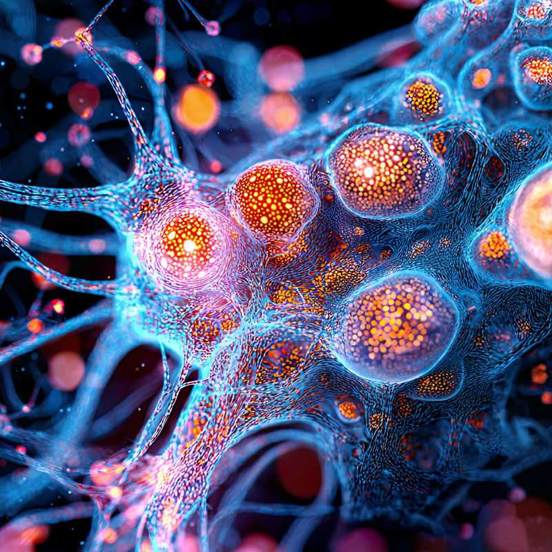
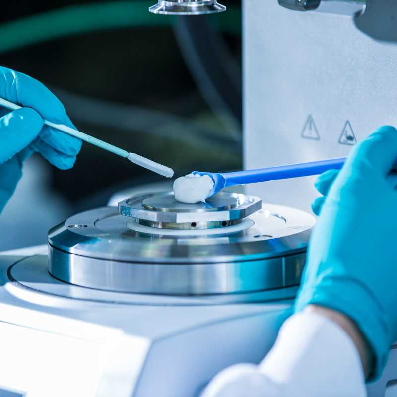
To accelerate your hit-to-lead campaign, we utilise cryo-electron microscopy (cryo-EM) to provide you with 3D structures of your challenging drug targets in complex with your compounds of interest. Our specialised protein science labs produce high-quality fresh protein samples, suitable for direct use in in-house grid preparation for cryo-EM. With regular access to high-end cryo-EM microscopes and over 16 years of cumulative industry experience, our team uses our cryo-EM platform to help ensure you receive detailed structural insights to drive your drug discovery efforts forward. Partner with us to leverage our expertise and state-of-the-art technology for your most demanding projects.

Iterative cycles of protein optimisation and grid screening are typically required to deliver high-resolution EM structures. Our EM platform provides access to fast cycle times, including:

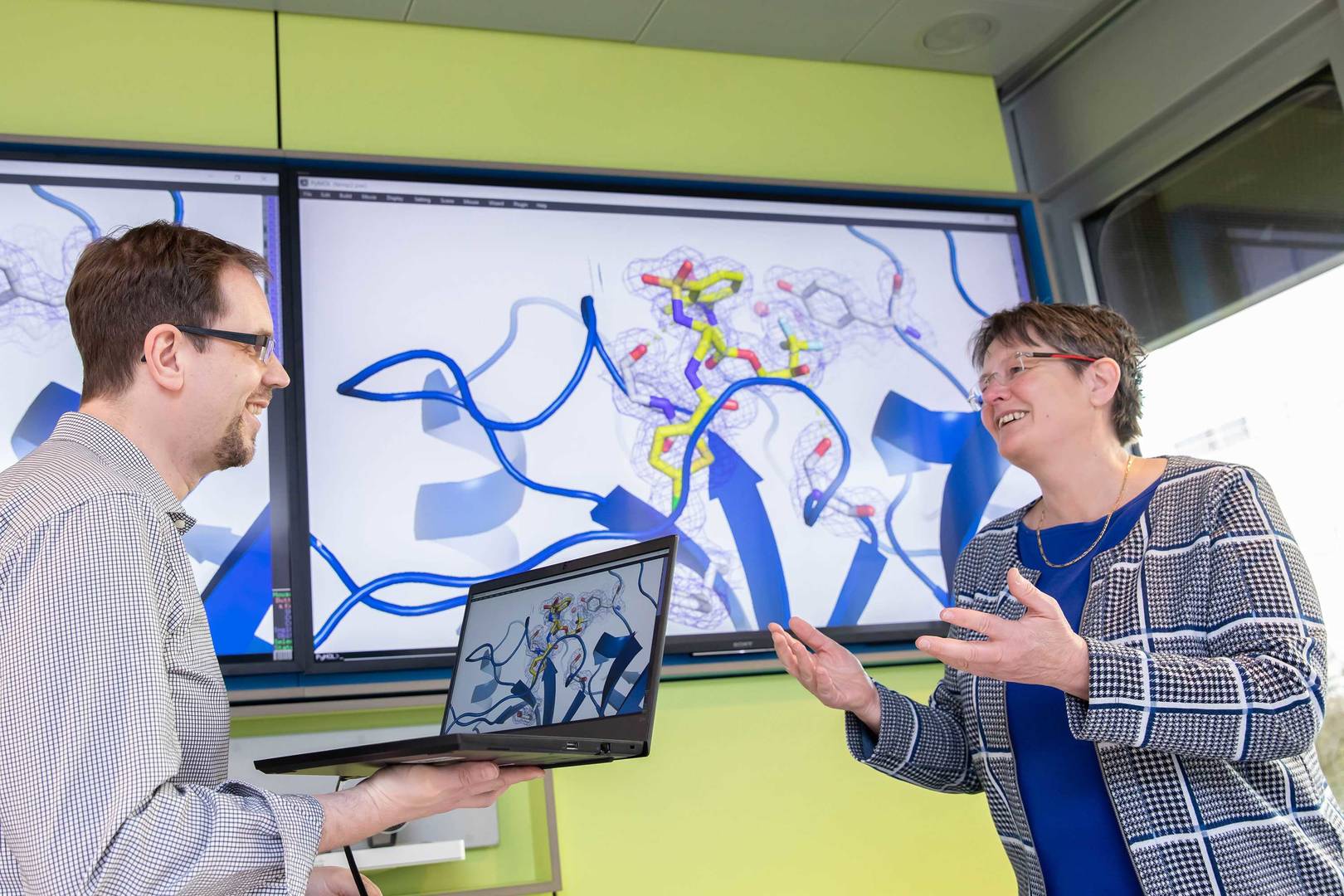
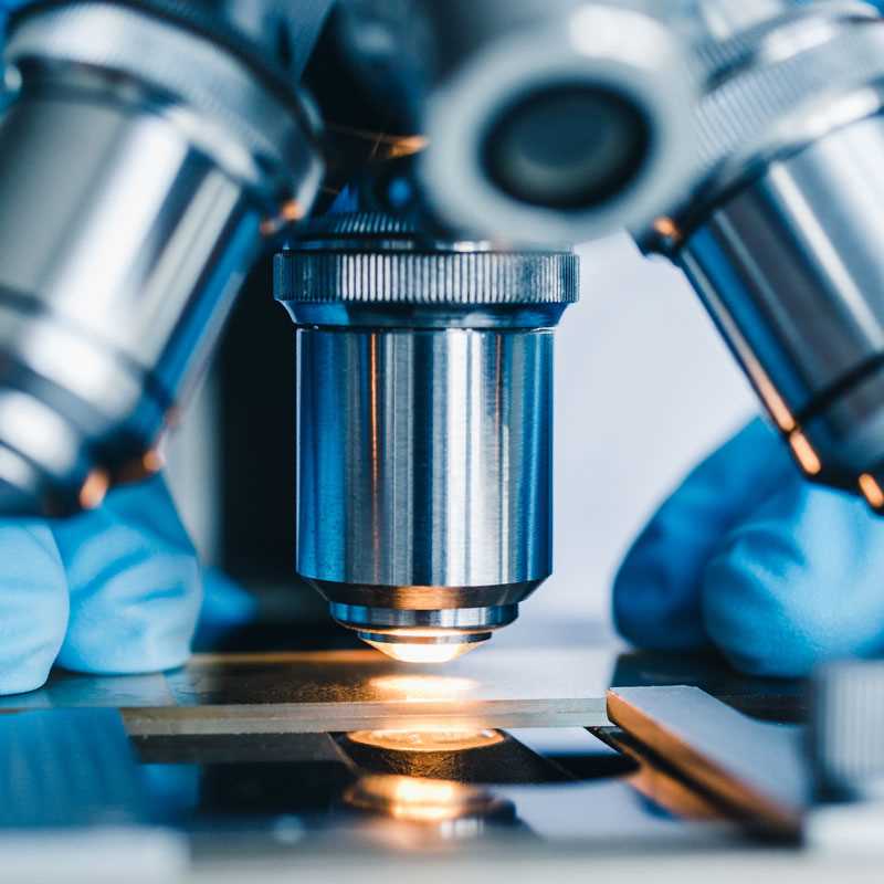
Nuvisan´s X-ray crystallography solutions offer detailed insights into molecular structures, aiding in drug design and development.
learn more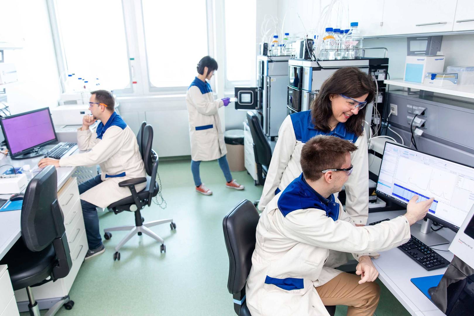
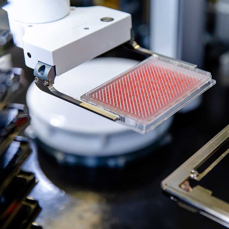
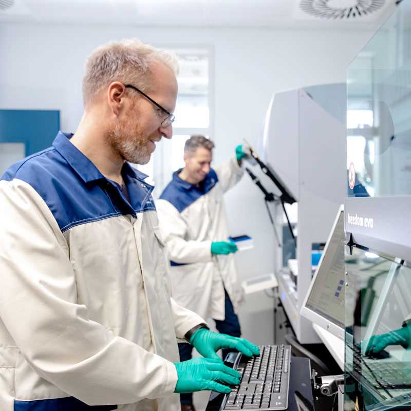
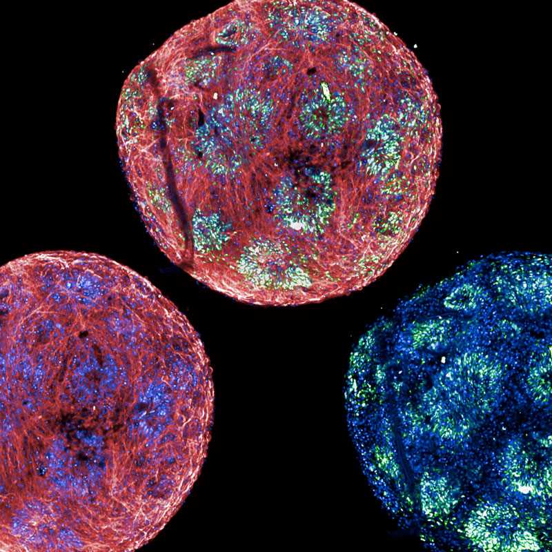
Our protein sciences team specialises in producing high-quality proteins essential for your research and development needs.
learn more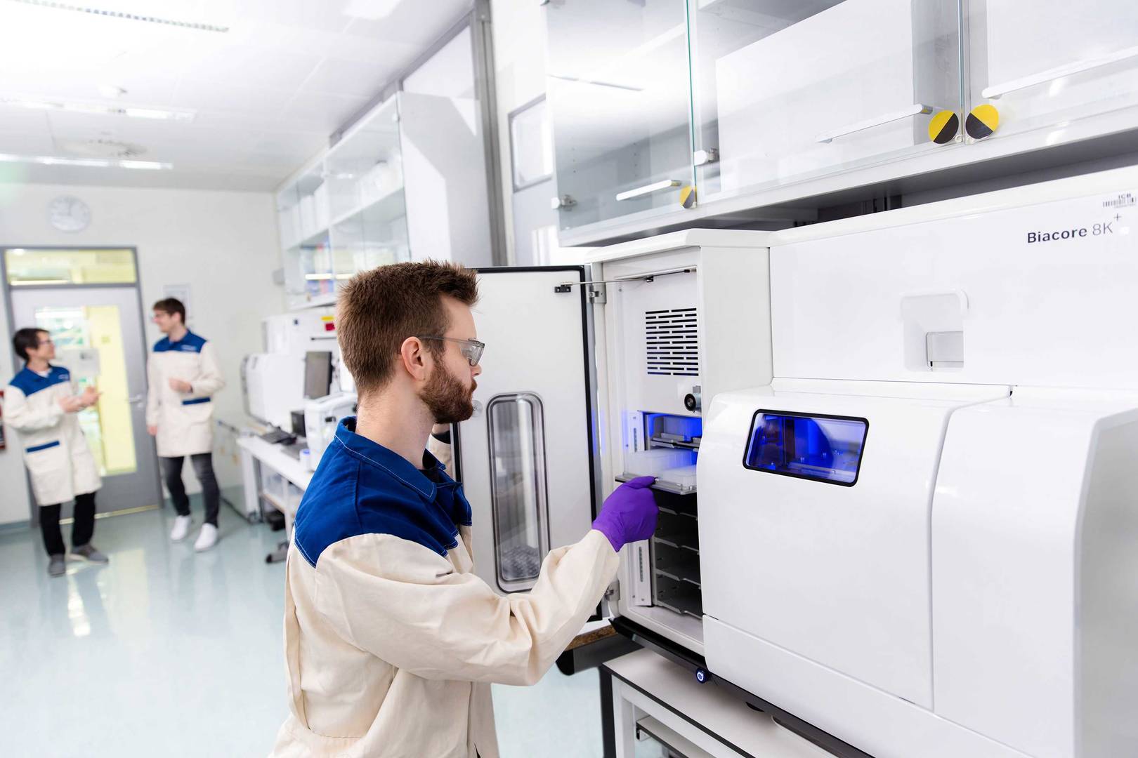
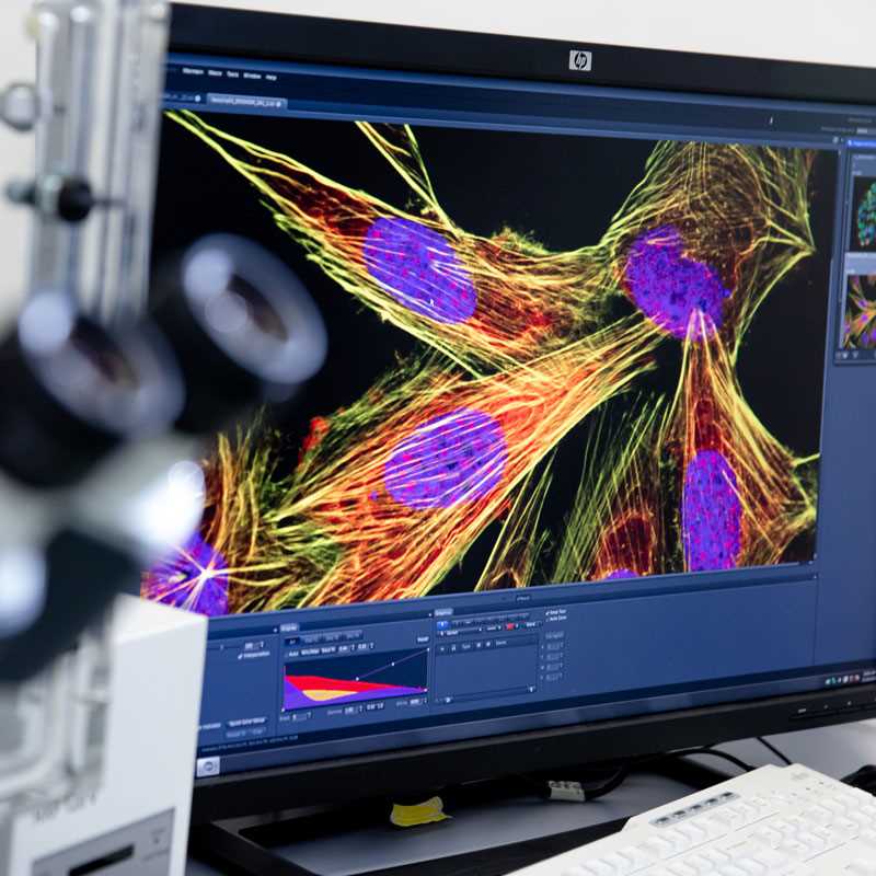
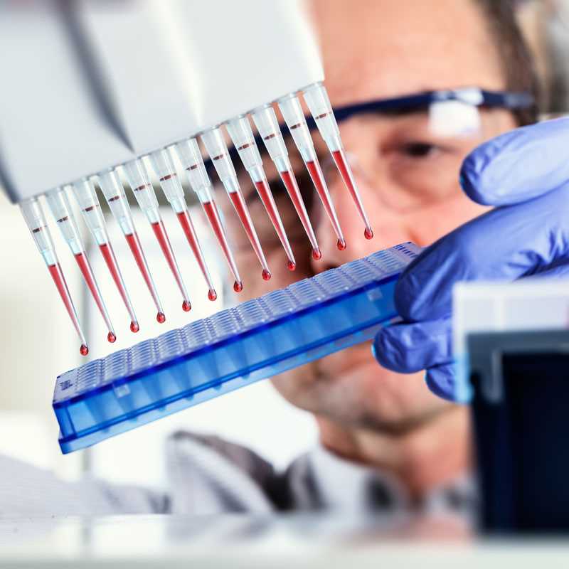
Our biophysical methods help validate interactions and optimise drug candidates.
learn more